Wu Laboratory

In the Wu Lab, we work to study and document the structures of neural systems at the whole organ and organism levels. Our study is guided by the questions:

“How do diverse neural circuits precisely form during development?”
&
“How are neural circuits disrupted by genetic mutations or disease?”

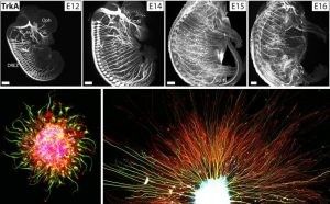
It is commonly known that complex neural circuits form within the brain to control basic biological processes and behavior. Similarly, neurons’ axons innervate varied organs in the peripheral nervous system, carrying signals that regulate the organs’ physiological functions. Understanding the architecture of these neural structures is important to understanding their function. Yet capturing a complete architecture poses unique challenges. When a single neuron’s numerous, sub-micron thin projections can extend across the entire brain and beyond in multiple directions, how can that neuron and its associated circuitry be investigated in detail in its entirety?

To tackle this challenge, we develop and apply novel three-dimensional imaging methods in our lab (the iDISCO family of tissue clearing) which we combine with genetic approaches. This platform allows us to image intact organs and embryos to examine their internal neural structures in true three dimensions. With this platform, we study how neural circuits assemble in the developing mouse embryo. This model helps us answer questions about how healthy circuitry is established and how neural wiring can go wrong. We also study adult animals to characterize the fully established circuitry and to investigate how ongoing changes in circuitry structure may play a role in aging and disease processes. Understanding the structural changes that occur will hopefully give us insights into how we may restore function in human individuals suffering from genetic mutations or disease.

We are a vibrant, inclusive, and synergistic team with multidisciplinary expertise (genetics and modern histology, cellular and molecular biology, advanced volumetric imaging, and big-data analysis using machine learning), to facilitate our highly collaborative research spanning basic systematic profiling of the brain to pathological discovery with animal models and human patient samples. Our motivation is to contribute to the development of both tools/resources and biological insights, as well as to engage in community effort for the education and training of scientists to lead future brain research.

Lab Projects

Whole Mount Development

whole mount clearing

Whole mount clearing

The spatial properties of neural structures—long, thin, and complexly oriented in three-dimensional space—pose unique challenges to imaging. Capturing these structures in their entirety requires high spatial resolution imaging through large depths and fields of view. Restricted by these limitations, traditional approaches slice organs into two-dimensional cross-sections to examine their interior.

Over the past seven years, we have developed novel whole mount imaging techniques to see inside organs without cutting or deforming their three-dimensional structure. Pairing tissue clearing—the process of making a tissue transparent—with immunohistochemistry, we label proteins of interest inside intact tissue to understand the identity, distribution, and spatial arrangement of both neuronal and non-neuronal cells. We image these cleared tissues using lightsheet microscopy. Our foundational method, iDISCO, was developed in 2014 for imaging intact embryos and brains. We have since continued to further develop our method, releasing iDISCO+ in 2016, and to optimize our approach for application to various peripheral organs, including adipose tissues which are traditionally difficult to clear due to their high lipid content (see AdipoClear, 2018).

We apply this technique to study a range of biological questions, both in our lab and with collaborators. The iDISCO family of approaches is not only useful for addressing questions about extensive, complex structures, such as neurons or vasculature, but also offers distinct advantages when studying disease processes where the location of one’s target is not known a priori by facilitating an unbiased survey of the bulk tissue.

3D Proteomics

3D proteomics

The mammalian brain contains numerous diverse cell types and functional states, which may undergo changes during disease processes. We are developing a three-dimensional proteomic database of the mouse brain captured at cellular resolution to provide fundamental knowledge of cell type distributions and structural morphology in the healthy adult mouse. These proteomic profiles will link new knowledge generated by single cell transcriptomic profiling to functional analyses.   To build this reference, we have established an efficient IHC pipeline to screen monoclonal antibodies (mAbs) for compatibility with whole-mount labeling and imaging based on our advanced iDISCO methods. We are building our reference using monoclonal antibodies because of the advantages these reagents provide in consistent quality and reproducibility. Employing these antibodies, we generate 2D and 3D datasets through whole mount imaging.

Our purpose in this project is three-fold: 1) To provide 3D whole mount proteomic datasets which can directly be used by other scientists to facilitate versatile molecular analyses of specific brain structures. 2) To build a reliable mAb reference list for numerous protein targets, to contribute to knowledge of reliable reagents and reproducibility within the field. 3) To disseminate optimized protocols with reference IHC results to direct the successful utilization of each mAb clone.

By sharing our resulting proteomic datasets on our publicly-accessible online platform (coming soon), we hope our reference can provide a valuable resource for the field.

Axon Guidance in Neurodevelopment

axon guidance

Axon guidance in neurodevelopment

We study how neural axon guidance establishes properly-formed neural circuitry in the developing mouse embryo. We use both in vivo genetics and in vitro culture models of embryonic neurons to investigate the mechanisms by which extending axons are able to navigate precisely in the complex environment and reach their proper targets over long distances. We are interested in understanding how myriad environmental cues (canonical guidance molecules and other ECM factors) orchestrate the dynamic axonal responses to form diverse neural projections. Studying this process will not only help us to understand the blueprint of adult neural circuitry formation to ensure mature functions, but also provide critical insights for mechanisms underlying later axonal plasticity, pruning and degeneration during aging and disease.

Weill Cornell Medicine Helen & Robert Appel Alzheimer’s Disease Research Institute 413 E. 69th St. New York, NY 10021